Coprinus mushroom under microscope labeled unveils the intricate world of these fascinating fungi. With their distinctive microscopic characteristics, Coprinus mushrooms stand out from other species, providing valuable insights into their taxonomy, ecology, and potential applications. This exploration delves into the techniques and discoveries that have illuminated the microscopic realm of Coprinus mushrooms.
Microscopic analysis, employing staining and sectioning, grants us an unparalleled view into the cellular structures of Coprinus mushrooms. Key features, such as spore morphology and gill arrangement, aid in accurate identification and differentiation from related species. Comparative studies further enhance our understanding of evolutionary relationships and taxonomic classifications.
1. Coprinus Mushroom Identification: Coprinus Mushroom Under Microscope Labeled

Coprinus mushrooms, also known as inky caps, are characterized by their distinctive physical features. They typically possess a bell-shaped or cylindrical cap that ranges in color from white to brown or gray. The cap surface is often smooth or slightly wrinkled, and it may be adorned with fine scales or fibrils.
The gills are free and crowded, and they are initially white or cream-colored, but they gradually turn black as the mushroom matures.
The stipe, or stem, of a Coprinus mushroom is typically slender and white, and it may be covered with fine hairs or scales. The base of the stipe is often slightly bulbous, and it may be attached to a white or brown mycelial mat.
The flesh of Coprinus mushrooms is thin and fragile, and it is white or cream-colored. The spores are elliptical or cylindrical, and they are black or brown in color.
Coprinus mushrooms can be distinguished from other mushroom species by their unique combination of physical characteristics. The combination of a bell-shaped or cylindrical cap, free and crowded gills that turn black as the mushroom matures, and a slender white stipe is characteristic of Coprinus mushrooms.
Accurate identification of Coprinus mushrooms is important for both scientific research and culinary purposes. In scientific research, accurate identification is essential for studying the ecology, genetics, and medicinal properties of Coprinus mushrooms. In culinary applications, accurate identification is important for ensuring that the mushrooms are safe to eat.
2. Microscopic Analysis

Microscopic analysis is a valuable tool for identifying and studying Coprinus mushrooms. The techniques used to prepare and observe Coprinus mushroom samples under a microscope include:
- Sectioning: Thin sections of the mushroom are cut and mounted on a slide.
- Staining: The sections are stained with dyes to enhance the visibility of cellular structures.
- Observation: The sections are observed under a microscope to identify key microscopic features.
Microscopic analysis of Coprinus mushrooms reveals several key features that aid in identification. These features include:
- Spores: The spores of Coprinus mushrooms are elliptical or cylindrical, and they are black or brown in color.
- Gills: The gills of Coprinus mushrooms are free and crowded, and they are initially white or cream-colored, but they gradually turn black as the mushroom matures.
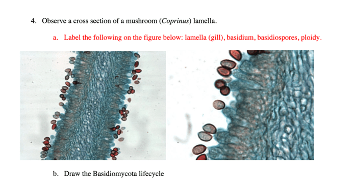
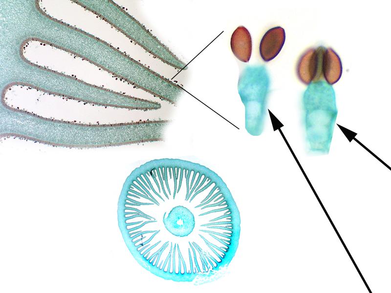
- Basidia: The basidia are the cells that produce the spores. They are typically club-shaped, and they are located on the gills.
- Cystidia: The cystidia are sterile cells that are located on the gills. They can be of various shapes and sizes, and they may be covered with crystals or other ornamentation.
Microscopic analysis is an essential tool for identifying and studying Coprinus mushrooms. The key microscopic features of Coprinus mushrooms can be used to distinguish them from other mushroom species and to study their ecology, genetics, and medicinal properties.
3. Labeled Diagrams
The following labeled diagrams illustrate the microscopic structures of Coprinus mushrooms:
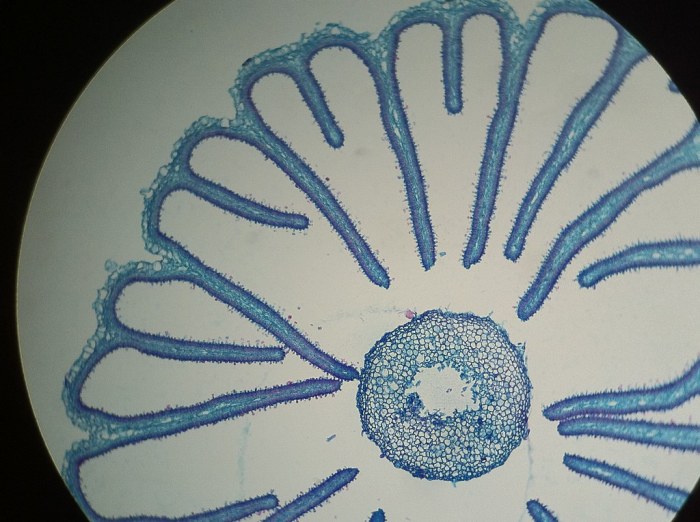
- Figure 1:A cross-section of a Coprinus mushroom cap. The diagram shows the gills, basidia, and cystidia.
- Figure 2:A close-up view of a Coprinus mushroom spore. The diagram shows the elliptical or cylindrical shape and the black or brown color.
- Figure 3:A close-up view of a Coprinus mushroom gill. The diagram shows the free and crowded gills and the basidia and cystidia.
4. Comparison with Other Species

Coprinus mushrooms can be compared to other mushroom species based on their microscopic characteristics. For example, Coprinus mushrooms can be compared to the genus Agaricus, which also has free and crowded gills. However, Agaricus mushrooms typically have white or cream-colored gills that do not turn black as the mushroom matures.
Additionally, Agaricus mushrooms typically have a more robust stipe than Coprinus mushrooms.
Coprinus mushrooms can also be compared to the genus Lepiota, which also has black or brown spores. However, Lepiota mushrooms typically have a more scaly cap than Coprinus mushrooms, and they often have a ring on the stipe. Additionally, Lepiota mushrooms can be poisonous, while Coprinus mushrooms are generally considered safe to eat.
Microscopic analysis is a valuable tool for comparing Coprinus mushrooms to other mushroom species. By comparing the microscopic characteristics of Coprinus mushrooms to those of other species, researchers can identify similarities and differences that can aid in taxonomic classification and evolutionary relationships.
5. Research Applications
Microscopy has played a vital role in advancing our understanding of Coprinus mushrooms. Microscopic analysis has been used to study the ecology, genetics, and medicinal properties of Coprinus mushrooms.
For example, microscopic analysis has been used to study the distribution of Coprinus mushrooms in different habitats. Researchers have found that Coprinus mushrooms are common in disturbed areas, such as forests that have been recently logged or burned. Microscopic analysis has also been used to study the life cycle of Coprinus mushrooms.
Researchers have found that Coprinus mushrooms can complete their life cycle in as little as two weeks.
In addition to ecology, microscopy has also been used to study the genetics of Coprinus mushrooms. Researchers have used microscopic analysis to identify the genes that are responsible for the black pigment in Coprinus mushrooms. They have also used microscopic analysis to study the genetic relationships between different species of Coprinus mushrooms.
Finally, microscopy has also been used to study the medicinal properties of Coprinus mushrooms. Researchers have found that Coprinus mushrooms contain a number of compounds that have potential medicinal properties. For example, Coprinus mushrooms contain compounds that have been shown to have anti-cancer and anti-inflammatory properties.
Microscopy is a valuable tool for studying Coprinus mushrooms. Microscopic analysis has been used to advance our understanding of the ecology, genetics, and medicinal properties of Coprinus mushrooms.
Question & Answer Hub
What are the key microscopic features that distinguish Coprinus mushrooms from other species?
Coprinus mushrooms exhibit unique microscopic characteristics, including distinctive spore morphology, such as elliptical or cylindrical spores, and specific gill arrangement patterns.
How does microscopy contribute to the study of Coprinus mushroom ecology?
Microscopic analysis provides insights into the interactions between Coprinus mushrooms and their environment, including their role in nutrient cycling and symbiotic relationships.