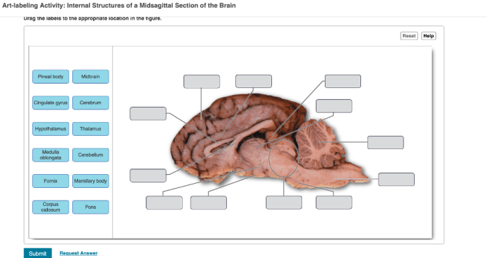
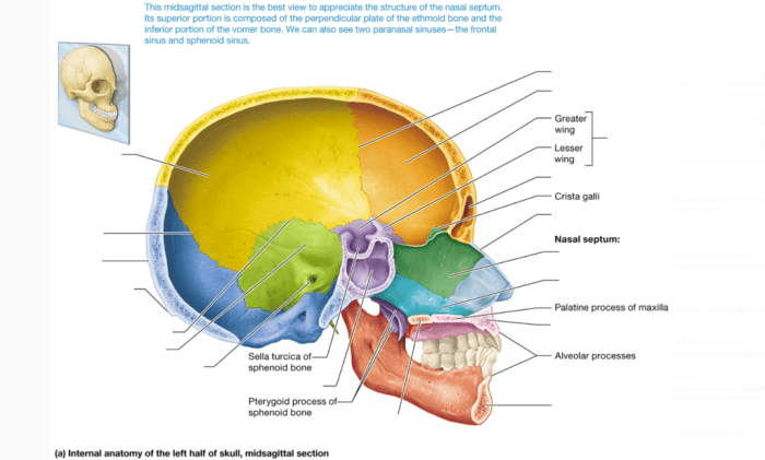
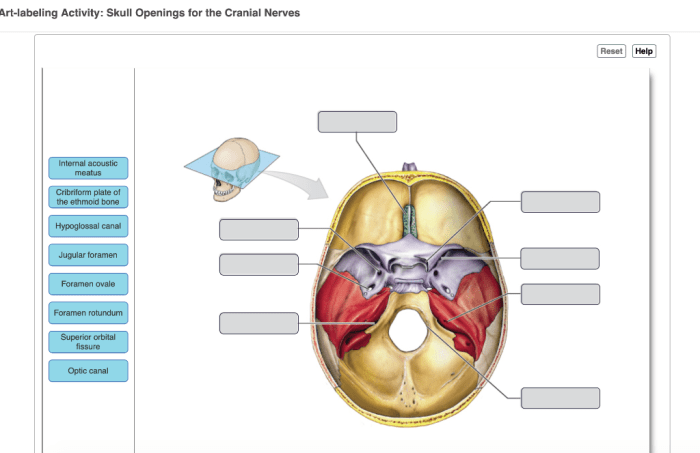
Art-labeling activity: internal midsagittal view of the skull provides a unique and engaging way to learn about the anatomy of the skull. This activity can be used in a variety of educational settings, from classrooms to museums. By labeling the different anatomical structures visible in the internal midsagittal view of the skull, students can gain a deeper understanding of the skull’s anatomy and how it functions.
The internal midsagittal view of the skull is a sagittal section that divides the skull into left and right halves. This view provides a clear view of the skull’s internal structures, including the brain, nasal cavity, and oral cavity. The internal midsagittal view is often used in medical imaging, such as CT scans and MRIs, to diagnose and treat skull injuries and diseases.
Art-Labeling Activity: Internal Midsagittal View of the Skull

The art-labeling activity is an educational technique that combines artistic expression with scientific learning. In this activity, students create a labeled diagram of an anatomical structure, such as the internal midsagittal view of the skull. By combining visual art with scientific labeling, this activity enhances student engagement and comprehension of complex anatomical structures.
Purpose and Objectives of the Art-Labeling Activity, Art-labeling activity: internal midsagittal view of the skull
The art-labeling activity serves multiple purposes and objectives in educational settings:
- Enhances Visual Learning:By creating a visual representation of the anatomical structure, students can better visualize and understand its three-dimensional relationships and spatial orientation.
- Improves Memory and Recall:The process of labeling and annotating the diagram helps students encode and retain information more effectively.
- Promotes Critical Thinking and Analysis:Students must carefully examine the anatomical structure, identify its key features, and determine appropriate labels.
- Encourages Creativity and Artistic Expression:The activity allows students to express their creativity through drawing and labeling, fostering a multi-sensory learning experience.
Identify the Internal Midsagittal View of the Skull
The internal midsagittal view of the skull is a cross-sectional view that divides the skull into equal right and left halves. This view provides a comprehensive view of the internal structures of the skull, including the:
- Frontal bone
- Parietal bone
- Occipital bone
- Sphenoid bone
- Ethmoid bone
- Maxillary bone
- Mandible
- Nasal cavity
- Pharyngeal cavity
- Oral cavity
- Pituitary gland
- Hypothalamus
- Cerebrum
- Cerebellum
- Brainstem
Key Questions Answered: Art-labeling Activity: Internal Midsagittal View Of The Skull
What is the purpose of the art-labeling activity: internal midsagittal view of the skull?
The purpose of the art-labeling activity: internal midsagittal view of the skull is to help students learn about the anatomy of the skull in a fun and engaging way.
What are the benefits of using the art-labeling activity: internal midsagittal view of the skull?
The benefits of using the art-labeling activity: internal midsagittal view of the skull include improved student understanding of skull anatomy, increased student engagement, and development of fine motor skills.
How can I use the art-labeling activity: internal midsagittal view of the skull in my classroom?
The art-labeling activity: internal midsagittal view of the skull can be used in a variety of educational settings, including classrooms, museums, and homeschool settings. The activity can be adapted to meet the needs of different students and can be used as a stand-alone activity or as part of a larger unit on skull anatomy.
